Computed tomography center
 International clinical trials submit high requirements for equipment of technological sites. Establishing diagnosis, determination of treatment strategy, online patient observation – all these are main principles of evidence based medicine.
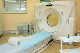
Our hospital is in effort to satisfy international standards. Buying computed tomography scanner Phillips Mx 8000 and specialists training was a significant stage in our direction.
Up-to-date computed tomography scanner Phillips helps to perform large variety of diagnostic procedures: non-invasive diagnostics (CT assessment), biopsy controlled with computed tomography etc…
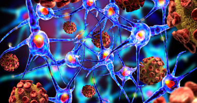
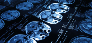
 Computed tomography – is the method of nondestructive examination of object internal structure. Method is based on measurement and complex computer processing of radiation attenuation difference in various issue texture.
During CT assessment radiologists evaluates a great set of images, and thus can see any abnormality. Slice thickness of 5 mm provides very detailed image, and helps to identify even smallest changes, defects or tumor formation. Furthermore, CT images can be combined with the help of computer and provide three-dimensional image of any body part (organ, vessel, bone).
CT assessment is much more convenient and informative than X-ray. To receive more exact data a contrast drug is injected.
Contrast drug is used to “light” some structures in images. Contrast drug blocks x-ray beams and light them 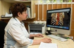 white, emphasizing blood vessels, intestines etc.. It enables doctor to determine abnormalities, which may not be seen without contrast. We use best contrast drugs such as Ultravist – 100 and Tmohexolum – 300 in out hospital. Patient is asked to drink contrast drug before assessment, and contrast drug is also infused during the assessment. Premedication drugs may be prescribed if it is necessary. Doctors pay attention to individual characteristics of each patient, medical history and concomitant abnormalities. Anesthesiologist controls contrast drug infusion and patient condition during the period of assessment. Patient safety is of high priority, therefore computed tomography center is equipped with emergency aid sets.
It should be noted that to perform objective evaluation of the disease in dynamics, to evaluate tumor response to treatment within the accuracy of decimals is only possible by comparing images made by the same scanner. Each patient participating in international clinical trials has the ability to perform this assessment for free and without waiting lines.
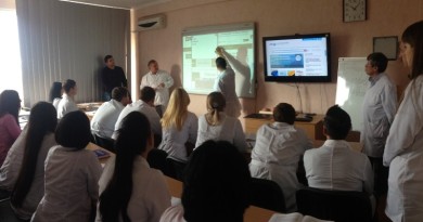
 Effective work of computed tomography center is ensured by specialists team.
Medical conclusions of assessments are given by radiologists of higher category. All center staff is trained and every time increases their qualification. A meeting with other specialists is performed in order to specify the diagnosis. We have implemented a remote work of radiologists. It became possible due to modern technology usage and high-speed internet.
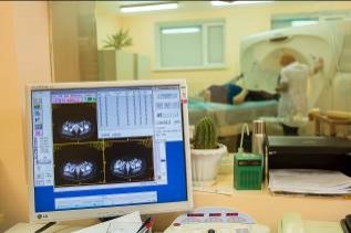
Data receiving and processing is provided by engineers and operator, who checks the equipment, performs diagnostic in timely manner and all other necessary measures required for effective work.
Special technical solutions helps to protect doctors and patients from radiation. Ventilation system, means of individual protection minimize health risks. High level of safety is approved by corresponding certificates and authorization documents.
Up-to-date diagnostic equipment, specialist team, patients’ safety and comfort – are the main component of computed tomography center. All this helps to diagnose diseases, consider any peculiarities in further treatment decisions, promptly respond in any changes, related to main disease. These steps allow to provide highly effective therapy, receive positive outcomes and improve patient quality of life.